Home
Uncategories
Rib Cage Muscles Labeled : 11 4 Identify The Skeletal Muscles And Give Their Origins Insertions Actions And Innervations Anatomy Physiology : Located in the rib cage, this muscle keeps the shoulder blade against the chest wall and helps rotate the shoulder blade upward.
Rib Cage Muscles Labeled : 11 4 Identify The Skeletal Muscles And Give Their Origins Insertions Actions And Innervations Anatomy Physiology : Located in the rib cage, this muscle keeps the shoulder blade against the chest wall and helps rotate the shoulder blade upward.
Rib Cage Muscles Labeled : 11 4 Identify The Skeletal Muscles And Give Their Origins Insertions Actions And Innervations Anatomy Physiology : Located in the rib cage, this muscle keeps the shoulder blade against the chest wall and helps rotate the shoulder blade upward.. The thoracic cage is a component of the thoracic wall and encloses the majority of the structures of the respiratory system. Woman stomach anatomy 7 photos of the woman stomach anatomy activate javascript anatomy of a woman body, female abdomen anatomy, female organ anatomy, human stomach anatomy, stomach anatomy and physiology, stomach anatomy antrum, stomach anatomy pictures, womens stomach anatomy, stomach, anatomy of a woman body, female. Perform dumbbell pullovers to work the muscles along your rib cage. The thoracic cage (rib cage) is the skeleton of the thoracic wall. Serratus posterior and pectoralis minor muscles assist with raising the upper ribs.
Don't just draw a generic rib cage shape in there. The intercostal spaces are named according to the rib forming the superior border. The sternocleidomastoid muscle, which comes from the jaw and crosses over the neck, moves the breastbone upward. It spreads out like a fan and covers the rib cage like an armor plate. Muscles that helpful in expanding the thoracic cavity are called the inspiratory muscles.
Thoracic Wall And Breast Illustrations from www.imaios.com Our ribcage exists to protect the heart and lungs. Try to be as accurate as you can with them. Human anatomy muscles rib cage, muscle anatomy rib cage, muscle rib cage pain, muscular anatomy of the rib cage, human muscles, human anatomy muscles rib cage, muscle. Scalene and latissimus dorsi muscles in the upper back also assist with raising the shoulder blade to add extra space in the rib cage. Human anatomy · april 18, 2021. Perform dumbbell pullovers to work the muscles along your rib cage. The first seven ribs in the rib cage are attached to the sternum by pliable cartilages called costal cartilages; Anatomy of the rib cage diagram in this image, you will find thoracic vertebrum, costochondral joint, costal cartilage, costal margin, costal arch, thoracic vertebrum, xiphoid process, xiphisternal joint, body, manubrial sternal joint, manubrium, the sternal notch in it.
Perform dumbbell pullovers to work the muscles along your rib cage.
This muscle helps rotate the upper arm. The thoracic cage presents with spaces between adjacent ribs, which are called intercostal spaces. Get all the information about pain under ribs in this article. The ribs protect vital organs within the thoracic cage, and they also assist with breathing. Rib bone anatomy quiz for students taking anatomy and physiology! With the upper ribs, closer to the nodule (and in the case of lower ribs, a little further from the nodule) they are curved and have a rough surface that connects them with muscles, angulus costae. The pectoralis major has two anatomic sections or heads: The thoracic cage (rib cage) is the skeleton of the thoracic wall. Anatomy of the rib cage diagram in this image, you will find thoracic vertebrum, costochondral joint, costal cartilage, costal margin, costal arch, thoracic vertebrum, xiphoid process, xiphisternal joint, body, manubrial sternal joint, manubrium, the sternal notch in it. Each are symmetrically paired on a right and left side. See more ideas about anatomy reference, anatomy drawing, human anatomy. The human rib cage is made up of 12 paired rib bones; The major abdominal muscles include the transverse abdominals, the rectus abdominis, and the external and internal oblique muscles.
Human anatomy muscles rib cage, muscle anatomy rib cage, muscle rib cage pain, muscular anatomy of the rib cage, human muscles, human anatomy muscles rib cage, muscle. Muscle spasms felt within the rib cage may also be caused by the abdominal muscles. These muscles connect the lower part of the spine to the ilium and the femur and. But there are other causes which could be serious and require a prompt medical care. The thoracic cage takes the form of a domed bird cage with the horizontal bars formed by ribs and costal cartilages.
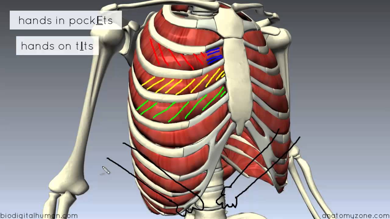
Muscles Of The Thoracic Wall 3d Anatomy Tutorial Youtube from i.ytimg.com The rib cage intrinsically holds the muscles of respiration (diaphragm, intercostal muscles, etc.) that are crucial for active inhalation and forced exhalation, and therefore has a major ventilatory function in the respiratory system. The remainder of the rib is the body of the rib (shaft). Scalene and latissimus dorsi muscles in the upper back also assist with raising the shoulder blade to add extra space in the rib cage. Of the remaining five ribs, which are called false, the first three have their costal cartilages connected to the cartilage above them. With the upper ribs, closer to the nodule (and in the case of lower ribs, a little further from the nodule) they are curved and have a rough surface that connects them with muscles, angulus costae. It is supported by the vertical sternum or breastbone (anteriorly) and the 12 thoracic vertebrae. The best way to learn anatomy is to repeat as much as you can. The major abdominal muscles include the transverse abdominals, the rectus abdominis, and the external and internal oblique muscles.
The rib cage intrinsically holds the muscles of respiration (diaphragm, intercostal muscles, etc.) that are crucial for active inhalation and forced exhalation, and therefore has a major ventilatory function in the respiratory system.
Human anatomy muscles rib cage, muscle anatomy rib cage, muscle rib cage pain, muscular anatomy of the rib cage, human muscles, human anatomy muscles rib cage, muscle. Construct a robo skelly rib cage and the pelvis using the bucket method. See more ideas about anatomy reference, anatomy drawing, human anatomy. The thoracic cage takes the form of a domed bird cage with the horizontal bars formed by ribs and costal cartilages. The thoracic cage (rib cage) is the skeleton of the thoracic wall. The human rib cage is made up of 12 paired rib bones; The upper edge is round and the lower sharp. This muscle helps rotate the upper arm. Look for clues from landmarks and muscle attachments that will tell you exactly where the rib cage is. Don't just draw a generic rib cage shape in there. Perform dumbbell pullovers to work the muscles along your rib cage. Anatomy of the body internal organs. Get all the information about pain under ribs in this article.
The body, or shaft, of the rib is thin, flat and curved. In the anatomical position, the angles align with the medial border of the scapula. Rib bone anatomy quiz for students taking anatomy and physiology! The rib cage intrinsically holds the muscles of respiration (diaphragm, intercostal muscles, etc.) that are crucial for active inhalation and forced exhalation, and therefore has a major ventilatory function in the respiratory system. Scalene and latissimus dorsi muscles in the upper back also assist with raising the shoulder blade to add extra space in the rib cage.
Rib Cage Labeled Page 4 Line 17qq Com from img.17qq.com Our most recent study sets focusing on rib cage muscles will help you get ahead by allowing you to study whenever and which of the. The recommended treatment for rib cage pain depends on the cause of the pain. Look for clues from landmarks and muscle attachments that will tell you exactly where the rib cage is. These muscles connect the lower part of the spine to the ilium and the femur and. These ribs are called true ribs. The remainder of the rib is the body of the rib (shaft). The major abdominal muscles include the transverse abdominals, the rectus abdominis, and the external and internal oblique muscles. The upper edge is round and the lower sharp.
This muscle helps rotate the upper arm.
The dome shaped thoracic cage provides the necessary rigidity for organ protection, weight support for the upper limbs and anchorage for muscles. The best way to learn anatomy is to repeat as much as you can. The body, or shaft, of the rib is thin, flat and curved. A rib has a flat body, as you can see from the picture of the anatomy of the human rib cage. A group of muscles connected to the rib cage, which help stabilize the shoulder. In this video, we explore:1) the anatomy of the sternum2) the anatomy and differences between the three classes of ribs3) the anatomy and differences between. The recommended treatment for rib cage pain depends on the cause of the pain. Introduction edit source. Woman stomach anatomy 7 photos of the woman stomach anatomy activate javascript anatomy of a woman body, female abdomen anatomy, female organ anatomy, human stomach anatomy, stomach anatomy and physiology, stomach anatomy antrum, stomach anatomy pictures, womens stomach anatomy, stomach, anatomy of a woman body, female. Muscles that helpful in expanding the thoracic cavity are called the inspiratory muscles. Located in the rib cage, this muscle keeps the shoulder blade against the chest wall and helps rotate the shoulder blade upward. These muscles connect the lower part of the spine to the ilium and the femur and. The thoracic cage takes the form of a domed bird cage with the horizontal bars formed by ribs and costal cartilages.
Post your work in the anatomy for artists rib cage muscles. Related posts of rib cage diagram with organs woman stomach anatomy.


0 Comments:
Posting Komentar